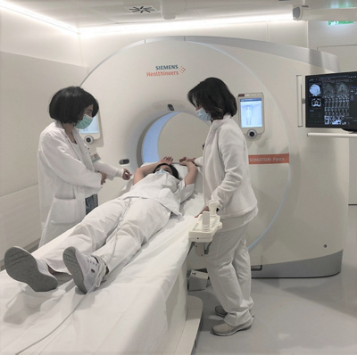
The Clinique des Grangettes benefits from state-of-the-art cardiovascular radiology equipment including, in addition to conventional equipment, an MRI and a scanner dedicated to cardiovascular activity.
Cardiovascular MRI
Cardiac magnetic resonance imaging (MRI) is a non-irradiating technique that provides dynamic images of the heart and clarifies the anatomy of this organ and the entire vascular tree of the body. Thanks to its ability to characterize the different tissues, this examination makes it possible to diagnose an inflammatory reaction, ischemia (suffering) or a scar of infarction. In addition, it provides an exhaustive and detailed analysis of the contours of the ventricles and volumes in three dimensions.
In practice, the examination takes place while lying on your back in the MRI machine. ECG electrodes are placed on the chest to synchronize image acquisition with the heartbeat. The examination requires the placement of a venous line, to inject either a contrast medium (called Gadolinium) or a drug to examine the heart under conditions of stress (pharmacological stress). In general, the acquisition of images is done during an apnea of a few seconds. The examination lasts between 30 and 50 minutes depending on the underlying pathology.
Cardiovascular CT scan
In the cardiovascular field, the scanner makes it possible to obtain images of thin sections of the heart and its arteries, all by using X-rays. This makes it possible to obtain a "virtual" coronary angiography of the heart or a "virtual" arteriogram of the arteries, all performed on an outpatient basis. Most of the time, the patient receives an intravenous injection of contrast medium in order to obtain more suitable images to make a more accurate diagnosis. The acquisition of images coupled with the use of an ECG lasts very little time and is therefore not too restrictive for the patient. The cardiovascular imaging specialist then analyzes the images obtained and thus makes the diagnosis of the patient concerned.


